Nuclear Medicine

Nuclear medicine is the use of radioactive substances for treating, diagnosing, and monitoring the effects of therapies for cancer patients, and investigating various other medical conditions including heart, and kidney disease. It can also be used to treat non-cancerous diseases such as hyperthyroidism. Both treatments and diagnoses involve administering radiopharmaceuticals - which are medicines that emit radiation.
How does it work?

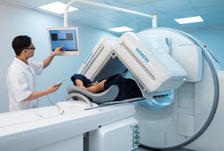
To diagnose disease, a range of different radiopharmaceuticals, each of which is preferentially taken up by a particular organ or type ofcell, can be injected into the bloodstream, swallowed or breathed in. The amount and distribution of radiopharmaceutical within the organ being studied is detected by a 'gamma camera', which may remain static or rotate right around the patient. Gamma cameras are usually linked to a computer, which uses image processing software to produce a map of the distribution of radioactivity as planar (two dimensional) images or as a series of sections through the body. Collecting a consecutive series of images will reveal how an organ is working.

For example, cardiac patients can be given radiopharmaceuticals to label their myocardium (cardiac muscle that forms part of the heart wall) so a moving picture of their beating heart can be obtained. Some tests begin as soon as the radiopharmaceutical has been administered, for example dynamic kidney imaging takes place immediately so the way the kidneys process the radiopharmaceutical can be followed.
By contrast, it takes several hours for the radiopharmaceutical used in bone scanning to leave the soft tissue surrounding the skeleton so that a clear picture of the amount of radiopharmaceutical that has been taken up by the bone can be obtained.
SPECT/CT (single photon emission computed tomography/computerised tomography) combines CT scanning - which is a series of X-rays taken at different angles through the body - with images captured via a gamma camera. The resulting cross-sectional images show both the structure (from the CT) and functionality (from the SPECT) of the area being studied.
In PET/CT (positron emission tomography/computerised tomography) imaging, the CT section of the scanner records a CT scan immediately before or after the PET scan. The PET portion of the scanner contains a circle of sensitive detectors that detect the radiation coming from the radiopharmaceutical that every patient is injected with before undergoing a scan. Diseased cells will absorb a different amount of this radiopharmaceutical than healthy cells, and so will be revealed in the scan by their different level of radioactivity. Cancer cells for example have a larger uptake than normal cells. 
Some diagnostic tests do not involve imaging. For example kidney function can be measured via blood and urine tests taken at particular time intervals after a radiopharmaceutical has been administered. For treatments, radiopharmaceuticals are swallowed or injected, and the energy from the radiation they give off destroys diseased cells. To treat thyroid cancer for example, radioactive iodine (I-131) in the form of either a liquid or a tablet is swallowed, then moves up into the neck to attack the cancer.
Who needs nuclear medicine?
Radiopharmaceuticals can be used to image patients with stress fractures, degenerative bone disease, or cancer patients with secondary cancer deposits in the bone, in addition to patients with lung, kidney, or gall bladder function problems. SPECT/CT is useful for investigating whether the heart muscle has the correct blood supply, and is commonly used to scan for certain types of tumour. Meanwhile, other patients with cancerous tumours including those in the uterus, thryroid, and prostrate are sometimes given radiopharmaceuticals as part of their treatment, as are some patients with non-cancerous conditions including hyperthyroidism and problems with the lubricating fluid around joints. PET/CT imaging is used to help diagnose and monitor treatments for patients with lung cancer, lymphomas and colorectal cancer, or to help diagnose neurological conditions such as epilepsy, and heart disease.
Future developments
PET/CT scanning is likely to become increasingly used in neurology and cardiology. Meanwhile one of the most recent radiopharmaceuticals developed can be used to help diagnose Parkinson's disease. In addition, gamma cameras that contain solid state radiation detectors similar to the light detectors used in digital cameras, are improving the resolution and sensitivity of the images obtained during many diagnostic tests